Exploring how the polyphenols in Morocco Gold extra virgin olive oil delivers the health benefits of bestolive oil – at a cellular level, beginning with polyphenol 3,4 DHPEA-EDA
Updated May 22nd 2023
Summary
- Morocco Gold extra virgin olive oil (EVOO) contains a range of polyphenols
- Polyphenols deliver the many health benefits associated with best olive oil
- Polyphenol 3,4 DHPEA-EDA in extra virgin olive oil (EVOO) acts as an anti-inflammatory and help protect the endothelium
- Damage to the endothelium and endothelial cells has now been linked with Covid-19
- The polyphenols in best olive oil like Morocco Gold extra virgin olive oil (EVOO) have been shown to reduce inflammation and oxidative stress in endothelial cells
Contents
- Factors Affecting Polyphenols In Morocco Gold Extra Virgin Olive Oil.
- Extraction Process For Extra Virgin Olive Oil
- The Polyphenols Present In Morocco Gold Extra Virgin Olive Oil
- Polyphenol : 3,4 DHPEA-EDA
- What This Polyphenol Does : It’s Role In Health Benefits
- The Endothelium, Blood Vessels and Endothelial Cells
- Where Do Endothelial Cells Come From And What Do They Do?
- Where Are Endothelial Cells Found?
- Heart, Brain, Liver, Lungs And Kidney
- Endothelial Cells And Cancer
- How Polyphenols In Extra Virgin Olive Oil Help To Protect The Endothelium
Factors Affecting Polyphenols In Morocco Gold Extra Virgin Olive Oil
The high polyphenol content of Morocco Gold extra virgin olive oil is dependent on three factors. First is the variety of the olive, secondly the climate and terroir of the growing region and thirdly the actual time in the growing season that the crop is harvested.
Morocco Gold is pressed from the Picholine Marocaine, the only type of olive to go into Morocco Gold extra virgin olive oil. Oil from this variety is renowned for its high polyphenol count, oxidative stability and longevity.
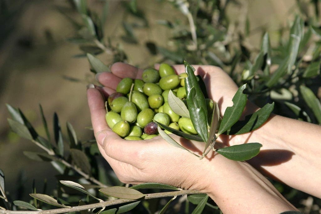
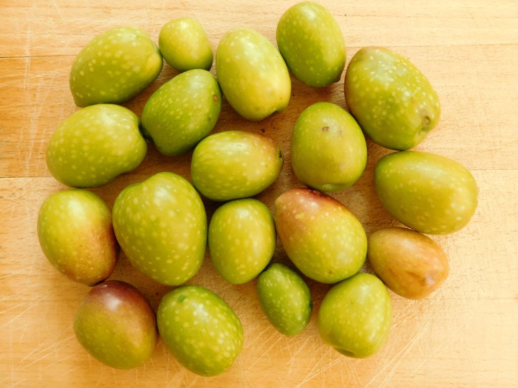
Thirdly, our olives are picked when the fruit is young and green. As the olives age on the tree, the colour of the olive changes to red and then black, the size of the olive increases thus producing more oil, but the polyphenol level decreases. There is a great deal of expertise within the farming community where we source our oil to ensure that the harvest is collected at the optimum time to maximise the polyphenol level in Morocco Gold extra virgin olive oil.
Extraction Process For Extra Virgin Olive Oil
The release of phenols and the formation of volatiles are two basic components of virgin olive oil quality that directly relate to the mechanical extraction process itself. In this ambit, control of endogenous enzymes of olive fruit during processing is the most critical point in the mechanical extraction process of olive oil. In fact, the secoiridoid concentration in the virgin olive oil is largely due to the activation of the glycosidases of olive fruit that activate the formation of aglycon, while the oxidoreductases such as polyphenoloxidase (PPO) and peroxidase (POD) can catalyze their oxidation during the oil mechanical extraction process and subsequently trigger the autoxidation mechanism.
Paolo Amirante, Alistair G. Paice
Olives and Olive Oil in Health and Disease Prevention, 2010
So what does all this mean? The types of polyphenol present in extra virgin olive oil is influenced by the extraction / pressing process. To be extra virgin olive oil, the extraction needs to be done solely by mechanical means and at low temperatures. This has a direct effect on the resulting polyphenol content. There also must be no mixing or blending with other oils and no chemical additives. This is how the natural goodness and health enriching qualities of the extra virgin olive oil is preserved.
The Polyphenol Content In Morocco Gold Extra Virgin Olive Oil
We are delighted to say that this year’s harvest has produced a low acidity level of 0.2% together with the highest level of polyphenols yet seen in our extra virgin olive oil.
| 3,4 DHPEA-EDA | 85 mg/kg |
| Hydroxytyrosol | 5 mg/kg |
| Lignanes | 26 mg/kg |
| Ligstroside aglycone (p, HPEA-EA) | 20 mg/kg |
| Oleuropein aglycone (3,4 DHPEA-EA) | 71 mg/kg |
| Oleocanthal p, HPEA-EDA | 65 mg/kg |
| Tyrosol | 372 mg/kg |
| Polyphenols Total | 644 mg/kg |
This confirms the ongoing high quality of this rare, delicious tasting extra virgin olive oil from this wonderful new source, together with its health enhancing qualities from its high polyphenol count.
Polyphenol : 3,4 DHPEA-EDA
Polyphenol 3,4 DHPEA-EDA (3,4-dihydroxyphenylethanol-elenolic acid dialdehyde) is a natural antioxidant found in extra virgin olive oil. 3,4-DHPEA-EDA is a polyphenol in the family of Tyrosols. It is synonymous with 3,4-DHPEA-Elenolic acid Di-Aldehyde and Oleuropein-aglycone di-aldehyde. It’s chemical formula is: C17H20O6
What This Polyphenol Content Does : It’s Role In Health Benefits
This compound has been studied extensively and has been shown to have numerous health benefits, including its ability to protect endothelial cells. Endothelial cells line the inner walls of blood vessels and play a crucial role in regulating blood flow and preventing the formation of blood clots. When these cells become damaged, this can lead to a range of health problems, including heart disease. By protecting endothelial cells, polyphenol 3,4 DHPEA-EDA in extra virgin olive oil may help reduce the risk of these conditions.
Diets in which fat is significantly provided by extra virgin olive oil have been associated with a low incidence of cardiovascular diseases. In this study the anti-inflammatory effect of 3,4-DHPEA-EDA on the endothelium was examined.
This study was based on the production of the proinflammatory chemokine CCL2, following in vitro stimulation of primary human endothelial cells. Pre-treatment of cells with 3,4-DHPEA-EDA resulted in a dose dependent inhibition of CCL2 secretion.
The effect of 3,4-DHPEA-EDA on CCL2 expression was observed at the transcriptional level. Functional data have shown that 3,4-DHPEA-EDA diminished monocyte adhesion to HUVECs. These results point on the use of 3,4- DHPEA-EDA as a novel drug aimed to prevent or reduce inflammation of endothelium.
It’s just one of the many benefits of consuming the best olive oil available that is rich in polyphenols. If you’re looking for a natural way to promote cardiovascular health, consider incorporating extra virgin olive oil into your diet.
Anti-Inflammatory Effect of 3,4-DHPEA-EDA [2-(3,4) www.researchgate.net › publication › 225288529_Anti-In…
The Endothelium, Blood Vessels and Endothelial Cells
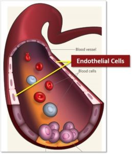
Almost all tissues depend on a blood supply, and the blood supply depends on endothelial cells, which form the linings of the blood vessels. Endothelial cells have a remarkable capacity to adjust their number and arrangement to suit local requirements. They create an adaptable life-support system, extending by cell migration into almost every region of the body.
Endothelial cells form the barrier between vessels and tissues. They control the flow of substances and fluid into and out of a tissue. An impaired function can lead to serious health issues throughout the body. Endothelial cells line blood vessels and lymphatic vessels, they are found exclusively in vascularized tissue.
Endothelial cells are nearly ubiquitous throughout the body. If it were not for endothelial cells extending and remodelling of the network of blood vessels, tissue growth and repair would be impossible.
Where Do Endothelial Cells Come From And What Do They Do?
Endothelial cells originate from the mesoderm, a germinal layer that forms at gastrulation, an early embryonic development stage (Bautch and Caron, 2015).
In medical research, endothelial cells are no longer seen as a passive barrier, but as a tissue that fulfils various functions (Michiels, 2003). These include numerous processes, such as blood vessel formation, coagulation and fibrinolysis, regulation of vascular tone, to a role in inflammation.
All endothelial cells share certain molecular characteristics: they test positive for von Willebrand factor (vWF), as well as for CD31 glycoprotein, and they test negative for smooth muscle alpha-actin.
This also applies to endothelial cells in capillaries. Here, endothelial cells are of tremendous importance for the exchange of substances including oxygen, water, and lipids, as well as in the transfer of carbon dioxide and urea from a tissue to a vessel, and vice versa. Depending on the tissue and organ, endothelial cells can vary from each other.
The soluble gas nitric oxide is essential for the regulation of the cells associated with blood vessel function (Tousoulis et al., 2012). Nitric oxide is a vasodilator and exerts its effects by relaxation of smooth muscle cells (Zhao et al., 2015). A dysfunctional NO-pathway is thought to be involved in cardiovascular disease.
Where Are Endothelial Cells Found?
Endothelial cells line all blood vessels. The largest blood vessels are arteries and veins, which have a thick, tough wall of connective tissue and many layers of smooth muscle cells. The wall is lined by an exceedingly thin single sheet of endothelial cells, the endothelium, separated from the surrounding outer layers by a basal lamina. The amounts of connective tissue and smooth muscle in the vessel wall vary according to the vessel’s diameter and function, but the endothelial lining is always present.
In the finest branches of the vascular tree, the capillaries and sinusoids, the walls consist of nothing but endothelial cells and a basal lamina, together with a few scattered but functionally important pericytes. These are cells of the connective-tissue family, related to vascular smooth muscle cells, that wrap themselves round the small vessels.
Heart, Brain, Liver, Lungs And Kidney
In the heart, three cell types enable the lifelong function of this continuously working organ: endothelial cells, fibroblasts, and cardiomyocytes (Lim et al., 2014). The communication between these cell types allows for organ development, autoregulation, and adaptation.
The most rigid separation between vessels and tissue can be found in the brain (Stamatovic et al., 2008). This structure is called the blood-brain barrier and the tight cell-to-cell junctions prevent most paracellular diffusion. Only substances with a specific transporter are picked up by the cerebral endothelial cells and are able to enter the brain.
At the other end of the spectrum, the most permeable endothelial cells of the mammalian body are located in the liver (Poisson et al., 2017). Liver sinusoidal endothelial cells build the interface between liver tissue and the portal vein originating in the gastrointestinal tract.
In the kidneys, endothelial cells play a key role in glomerular filtration (Satchell and Braet, 2009). Fenestrations in the endothelium allow for passage of molecules with a defined size. This prevents an excessive loss of proteins. This mechanism complements the filtration occurring through podocyte filtration slits.
To enable an efficient gas exchange in the lungs, the endothelial barrier must be intact (Rounds et al., 2008). Additionally, a prerequisite for optimal gas exchange is that the alveolar surface remains dry.
Endothelial Cells And Covid
Emerging scientific knowledge research suggests that Covid-19 attacks the body by causing inflammation and oxidative stress in the endothelial cells.
Cytokines are small proteins that are crucial in controlling the growth and activity of other immune system cells and blood cells. When released, they signal the immune system to do its job. Cytokines affect the growth of all blood cells and other cells that help the body’s immune and inflammation responses.
Although cytokines are an important component of our immune system, too many of these can lead to what is known as a ‘cytokine storm’. This is essentially an overreaction of our immune system which can have a seriously detrimental impact on COVID-19 sufferers and, in some cases, lead to fatality. This is because cytokine storms reduce the amount of oxygen circulating in our blood, causing fluid build-up in the lungs and lead to life-threatening breathing difficulties.
Endothelial Cells And Cancer
Cancerous tissue is as dependent on a blood supply as is normal tissue, and this has led to a surge of interest in endothelial cell biology. It is hoped that by blocking the formation of new blood vessels through drugs that act on endothelial cells, it may be possible to block the growth of cancerous tumours.
Endothelial cells are nearly ubiquitous throughout the body. However, there are two major instances where dysfunction of endothelial cells is involved in pathogenesis of a medical condition. First, in coronary artery disease, endothelial cells are damaged. So the generation of new vascular cells to restore organ function following a myocardial infarction is of high research interest. Understanding this process could help scientists to develop supportive myocardial therapies (He at al., 2017). Another example is atherosclerosis, where endothelial dysfunction arising from chronic inflammation within the arterial wall causes a pathological change of blood vessels (Gimbrone and García-Cardeña, 2016).
The endothelium is crucial for nutrient supply, which is why endothelial cells play an important role in cancer progression (Dudley, 2012). Tumors are defined by a high vascularization activity that can lead to irregular diameters, a fragile anatomy, and even a leaky structure. Understanding the biology behind these mechanisms and studying all factors and cell types involved could be one approach to developing new antiangiogenic therapies.
How Polyphenols In Extra Virgin Olive Oil Help To Protect The Endothelium
The antioxidant activity of polyphenols 3,4-DHPEA, p-HPEA and phenyl acids has been studied, and the high antioxidant activity of 3,4-DHPEA, 3,4-DHPEA-EDA and 3,4-DHPEA-EA has been demonstrated. Several nutritional properties have also been recognized:
- Inhibition of blood platelet aggregation and involvement in the synthesis of thromboxane in the human cells
- Inhibition of phospholipids and LDL oxidation
- Protection of the human erythrocytes from the oxidative damage.
Taken together, these factors indicate a role for polyphenols in extra virgin olive oil as a possible protective effect with respect to the risk of thrombosis and the onset of atherosclerotic damage in consumers.
Minor dietary constituents of extra virgin olive oil would seem to have major biochemical importance at the cellular level.